Soft robotic heart analog opens new possibilities for studying diseases
English translation from Unibot. Source published 2026-07-29.
UNSW researchers have created a soft robot that replicates the human heart. It is capable of simulating diseases and creating a realistic environment for testing new cardiac devices.
Breakthrough in cardiac modeling
Scientists at the University of New South Wales Sydney have developed a fully synthetic soft robotic heart. It replicates the complex movements and internal structures of a real organ - paving the way to better treatments, safer medical devices and personalized medicine.
The results were published in the journals Nature Communications and Advanced Science. The work presents a beating model of the left side of the heart with artificial valves, papillary muscles and chordae - the very structures that are critical for healthy heart function and often affected by disease.
The device accurately reproduces the situation when the heart valves allow blood to flow back - this increases the risk of heart failure and other dangerous complications.
According to the team, the new soft robot will eventually help to better understand cardiac pathologies, reduce animal testing and give doctors personalized patient models to plan surgeries before they are performed.
Why is this important
Team leader Scientia Associate Professor Thanh Nho Do, from UNSW's School of Biomedical Engineering and Medical Robotics Laboratory, said the work is important because cardiovascular disease remains the world's leading cause of death.
“Heart failure with preserved ejection fraction (HFpEF) is a complex condition that often comes hand in hand with other problems: high blood pressure, arrhythmia, kidney disease, obesity and diabetes,” says Professor Do. “Because it affects everyone differently, developing medical devices to improve heart function is challenging.
The heart valves are critical to its effectiveness, but disease can cause them to become flabby or stiff. This increases the load on the heart and can lead to heart failure.
Our ultimate goal is to create realistic artificial heart models that will help researchers understand diseases and develop safer and more effective devices before they are tested in animals or used in patients."
How a beating heart was recreated
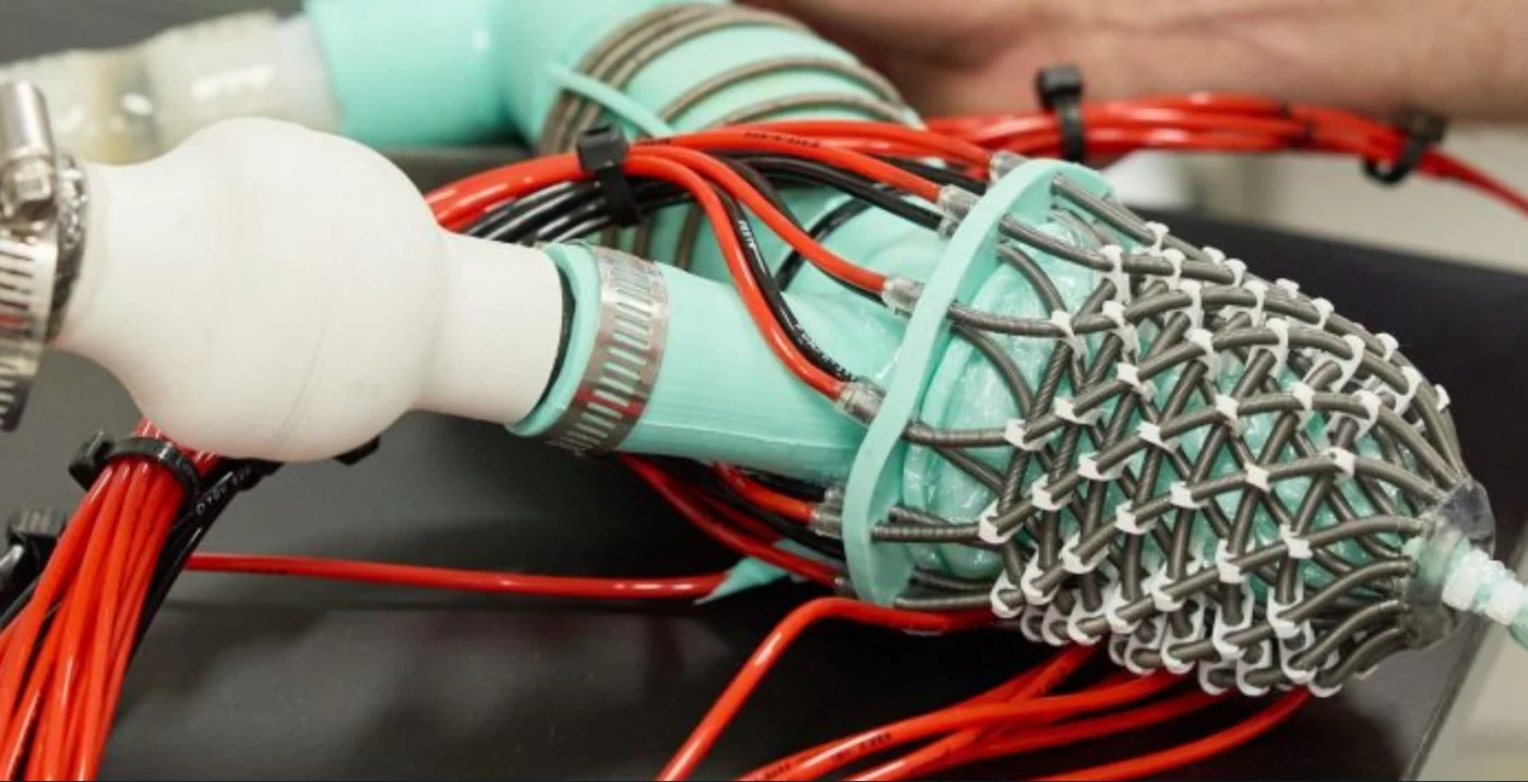
The model, developed at UNSW, is a soft, flexible replica of the left side of the heart. The internal chambers are made of silicone membranes, and soft robotic artificial muscles are wrapped around them - they reproduce the natural contraction and twisting of the organ.
Unlike conventional laboratory models, this heart contains structures that control the mitral valve. In reality, it works like a pair of swing doors: it opens and closes with each blow, so that oxygenated blood flows into the body and does not leak back. Thanks to this detail, researchers will be able to reproduce diseases in which the valve allows blood to flow backwards.
“The model is made of flexible materials and is driven by artificial muscles arranged to mimic the layered muscular architecture of the human heart,” explains Dr. James Davis, a postdoc in Do's group. “We have found a way to simulate this architecture using soft robotic artificial muscle fibers.” They work from hydraulic pressure, which we control - so our model of the ventricle moves like a real one.
We then wrap this artificial muscle around silicone membranes that mimic the inside of a human left heart. This is how we get our model of the left heart and atrioventricular apparatus. The membranes keep the simulated blood inside the left heart and allow it to be pumped into and out of the model.”
The system gives researchers the ability to actively adjust the tension of the artificial papillary muscles that support the mitral valve. By changing it, the team recreated the disease conditions mitral valve prolapse and regurgitation, where blood leaks backwards instead of moving efficiently through the heart.
Simulating human heart diseases
Using ultrasound and blood pressure measurements, scientists have shown that the artificial heart behaves strikingly similar to the real one. Healthy valves produced normal pressure and flow readings, and when the disease was simulated, the characteristic changes that doctors see in patients appeared.
“In the first study to replicate the internal valve system of the human heart, we were able to obtain pressure and blood flow curves that were close to real life,” says Professor Do. “Crucially, we have learned to regulate the function of the mitral valve by changing the length of the papillary muscles. We confirmed this with invasive measurements of pressure and flow inside and outside the heart, but also proved the model's compatibility with non-invasive clinical methods such as ultrasound or echocardiography. "The simulated healthy mitral valve met physiological expectations for pressure and flow, and in the disease simulation we observed increased regurgitation (backflow) and a drop in output pressure and flow - all consistent with real heart valve disease."
Professor Nigel Lovell, Head of the School of Biomedical Engineering and Director of Tyree IHealthE, added: “The ultrasound pattern was also human-like - thanks to the biomimetic shape and function of our model. "We saw the movement of the valve leaflets as in humans, and we observed blood flow through the valves, including the regurgitant jets that flowed from the valves in simulated disease."
The researchers also tested a new soft robotic cardiac catheter on a beating model. It was able to move inside an artificial heart and make contact with moving structures - showing how the platform could speed up the development of future surgical tools.
Less animal testing
Because the simulator provides a controlled and repeatable environment, scientists believe it will reduce the need for animal testing in the early stages of medical device development. “We hope to create a platform for comprehensive modeling of cardiac diseases and their treatments, from implants to surgical instruments,” says Professor Do. “Especially in the early stages of development, such a platform will allow us to monitor cardiac function while maintaining anatomical and physiological fidelity, and reduce the dependence on animal models, and with it the costs and ethical problems.”
By modeling a wide range of specific heart diseases, such as HFpEF, one of the most poorly understood and difficult to treat forms of heart failure, we hope to help create new implants and devices that save and improve lives by reducing the burden of cardiovascular disease on the healthcare system. HFpEF, which accounts for 50% of heart failure cases, deserves its own mechanical treatments.”
More importantly, the model successfully reproduced many of the changes characteristic of HFpEF, including disruptions in heart function and blood flow. When the researchers modeled one of the early signs of HFpEF—a decreased ability of the heart to relax between beats—the model showed that blood flowed to the heart more slowly and less efficiently. This delay in filling increased the pressure inside the heart - much like in patients with HFpEF.
The scientists also suggest that in the future it will be possible to create patient-specific versions of the model based on medical imaging data. Such personalized models will help physicians evaluate different devices and treatment approaches preoperatively, improving surgical planning and potentially outcomes.
“With the development of personalized medicine, we hope to provide better simulation of the cardiovascular system for a specific patient - this will help in planning operations and choosing the type, size and parameters of the implant,” says Professor Do. “We look forward to testing these concepts and moving toward clinical implementation.”
Future Validation
While the study demonstrates the potential of the technology, the scientists stress that the current model is more of a proof of concept than a ready-made clinical tool. A number of challenges remain: improving materials, refining control systems and making the device even more compatible with medical imaging. Future versions should better replicate certain aspects of the heart and use patient-specific geometries rather than simplified structures.
The most important thing is that the platform must be tested on real patient data. “The next key step is deeper validation on clinical data,” says Professor Do. - Current research demonstrates a strong evidence base for the concept. The model is capable of reproducing key parameters of pressure, blood flow, motion, valve function and imaging that correspond to the behavior of the human heart. But before this platform can be used for clinical decision making, it must be systematically compared with data from patients with a wide range of cardiac anatomy and disease severity.”
The team also includes Professor Christopher Hayward, a heart failure and transplant cardiologist at St Vincent's Hospital in Sydney, as well as Professor Elena Rnjak-Kovachina and Scientia Associate Professor Hoang-Phuong Phan from UNSW. The researchers hope that with further development of the technology, it will find application in clinical practice.
“We view the current model not as an off-the-shelf clinical tool, but as a platform that opens up new opportunities,” added Dr. Davis. “It shows that soft robotic artificial hearts can reproduce the mechanics of disease in a way that conventional laboratory models cannot.” And it provides a clear path to patient-specific modeling, device testing, and ultimately treatment planning.”
University of New South Wales